You are visiting a website that is not intended for your region

The page or information you have requested is intended for an audience outside the United States. By continuing to browse you confirm that you are a non-US resident requesting access to this page or information.

Switch to the US site

Select Your Country or Region
Continue
COVID-19 Resource Center

NAVA

patient and two nurses using the servo-u

NAVA

Ventilation where the patient’s own respiratory drive controls timing and assist delivered by the ventilator.

Overview

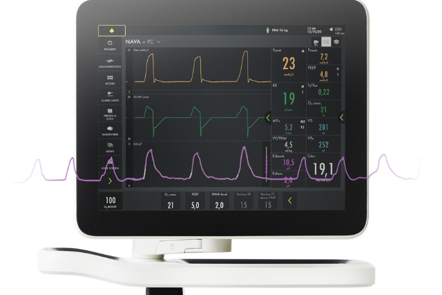
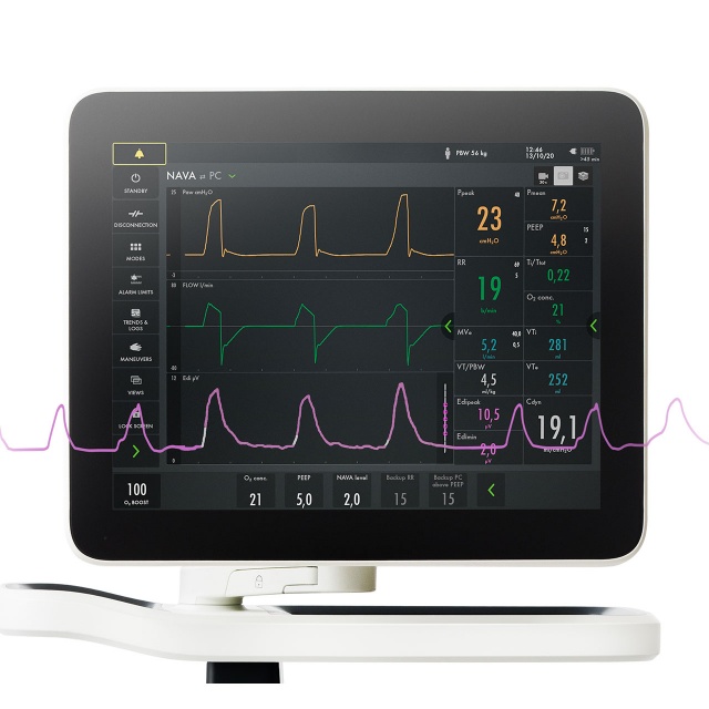
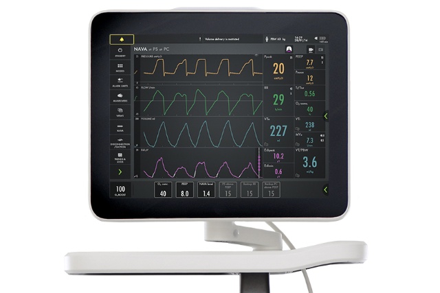
Personalized ventilation provides unique patient insight and ventilation capabilities. It consists of a diagnostic tool that helps you monitor diaphragm activity (Edi) on the ventilator screen and a ventilation mode (NAVA) that uses the diaphragm activity to deliver assist adapted to the patient.

Personalized ventilation can help you:

Support throughout the treatment

illustration nava

Invasive NAVA

Synchronized assist, weaning and sedation management, supporting early diaphragm activation.
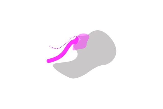
Illustation non-invasive nava

Non-invasive NAVA

Synchronized assist, independent of leakages allowing a gentler mask application.
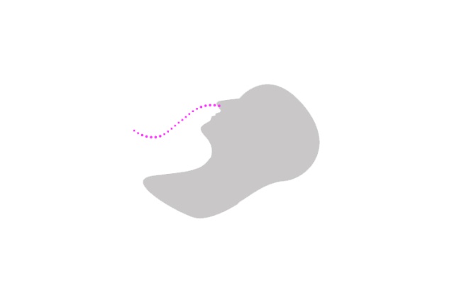
illustration monitoring

Edi monitoring

Monitor diaphragm activity  and breathing effort after extubation. Can be used with High Flow therapy if needed.


See and deliver what your patient wants

In most intensive care units 20% of patients consume 80% of ventilation resources, which may lead to increased complications and unwanted outcomes. [11] For these patients conventional ventilation simply isn’t enough. With personalized ventilation, the ventilator shows you what the patient wants, which may help you wean earlier with increased comfort, decreased sedation and reduced complications.

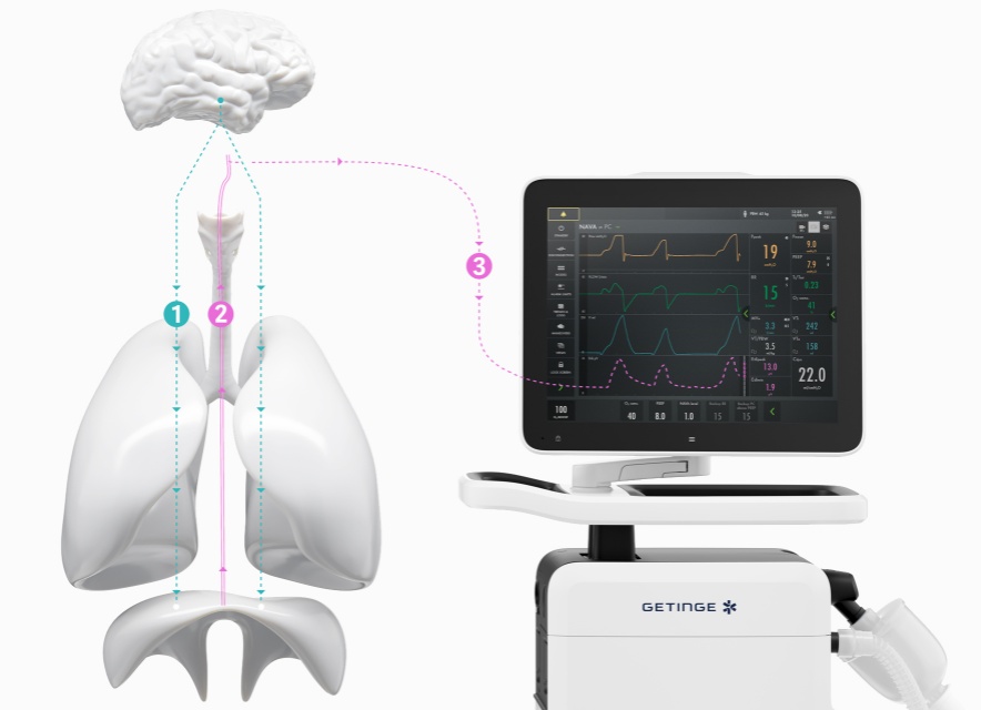
During normal respiration, a spontaneous breath begins with an impulse generated by the respiratory centers in the brain. This impulse is then transmitted via phrenic nerves and electrically activates the diaphragm, leading to a muscle contraction. The diaphragm contracts into the abdominal cavity, which leads to a descending movement, creating a negative alveolar pressure and an inflow of air.

The signal that excites the diaphragm is proportional to the integrated output of the respiratory center in the brain and controls the depth and cycling of the breath.

With personalized ventilation the electrical discharge of the diaphragm is captured by a special catheter fitted with an array of electrodes (the Edi catheter) and visualized on the ventilator screen. This is Edi, the electrical activity of the diaphragm. The Edi catheter is placed in the esophagus much like an ordinary feeding tube. With NAVA, Neurally Adjusted Ventilatory Assist, the Edi is used to deliver ventilation in time with and in proportion to the diaphragm activity.

1. Identify common ventilator challenges

Only 10% of experienced clinicians detect auto-triggering, one of many challenges that can result in patient-ventilator agitation, increased sedation and delayed weaning. This is because the ventilator waveforms show you what the ventilator delivers, not what the patient wants. [12]

Seeing patient diaphragm activity on screen (Edi) helps you:

  • monitor and safeguard the patient’s diaphragm activity [13] [14]
  • assess effort and work of breathing during weaning [15]
  • prevent muscular exhaustion during weaning trials, even after extubation. [16]

2. Keep the diaphragm active

Edi helps you detect diaphragm activity early and Neurally Adjusted Ventilatory Assist (NAVA) helps you exercise the diaphragm on a personalized level. [17] [18]

3. Protect the lung from injury and wean earlier

An active diaphragm is the first step towards successful weaning. The second step is to avoid lung injury. NAVA delivers assist in proportion to and in synchrony with the patient’s respiratory efforts, which can contribute to:

  • fewer periods of over- and under-assist [19] [20]
  • Improved patient-ventilator synchrony [12] [20]
  • reduced sedation [21] [22]
  • improved comfort scores [23]
  • improved sleep quality [24] [25]

The benefits of Edi monitoring

The diaphragm is the “heart” of the respiratory system and is designed to be continuously active. [26] The Edi is a bedside diagnostic tool that allows you to monitor and safeguard the patients diaphragm activity. [27] [28] The Edi guides weaning [29] and helps you prevent muscular exhaustion during weaning trials, even after extubation. [30]

How to detect an inactive diaphragm

Below notice how the pressure curve (yellow) to the middle and to the right seems just fine, even though the diaphragm as demonstrated by the Edi signal (pink) is inactive compared to the image on the left where the pressure curve follows the shape of the Edi signal.

graph

Active diaphragm (NAVA ventilation)

graph over-sedation

Over-sedation (Pressure support)

Graph over-assist

Over-assist (Pressure support)

How to detect patient ventilator-asynchrony

Identifying any of the many asynchronies is just as easy. Below are a few examples of asynchronies visible directly on the screen, continuously.

graph ventilator asynchrony

The benefits of NAVA ventilation

NAVA [31] follows the Edi, and allows the patient to select the tidal volume and respiratory pattern. As such NAVA promotes lung protective spontaneous breathing [32]  [33]  [34] with higher diaphragmatic efficiency, [35] [36]  and fewer periods of over and under-assist. [37]  [38] The patient’s ICU experience can be improved by reduced sedation, higher comfort scores [39]  [40]  [41] and improved sleep quality. [42]  [43] NAVA simply delivers what the patient wants.

Edi and NAVA assure that breathing efforts from all patient categories are effectively assessed and responded to. NIV NAVA is also independent of leakage in patient interfaces and may prevent respiratory failure and intubation. [44] [45] [46] 

Training

Improve your knowledge with our eLearning courses

The basic concept of NAVA and Edi
NAVA module 1 (10 min)

  • Breathing regulation
  • Conventional ventilatory treatment
  • Edi and NAVA treatment

English (voice over)

The basic concept of NAVA and Edi
NAVA module 2 (10 min)

  • Breathing regulation
  • Conventional ventilatory treatment
  • Edi and NAVA treatment

English (voice over)

Servo-u NAVA (10 min)

  • Servo-u NAVA screen layout
  • NAVA workflow

English (voice over) | French | German | Italian | Spanish | Swedish | Dutch

Downloads

All references

  1. Yonis H, et al. Patient-ventilator synchrony in Neurally Adjusted Ventilatory Assist (NAVA) and Pressure Support Ventilation (PSV). BMC Anesthesiol. 2015 Aug 8;15:117.

  2. Piquilloud L, et al. Neurally adjusted ventilatory assist improves patient-ventilator interaction. Intensive Care Med. 2011 Feb;37(2):263-71.

  3. Blankman P, et al. Ventilation distribution measured with EIT at varying levels of PS and NAVA in Patients with ALI. Intensive Care Med. 2013 Jun;39(6):1057-62.

  4. Patroniti N, et al. Respiratory pattern during neurally adjusted ventilatory assist in acute respiratory failure patients. Intensive Care Med. 2012 Feb;38(2):230-9.

  5. Kallio M, et al. Neurally adjusted ventilatory assist (NAVA) in pediatric intensive care – a randomized controlled trial. Pediatr Pulmonol. 2015 Jan;50(1):55-62.

  6. Piastra M, et al. Neurally adjusted ventilatory assist vs pressure support ventilation in infants recovering from severe acute respiratory distress syndrome: nested study. J Crit Care. 2014 Apr;29(2):312.e1-5.

  7. De la Oliva P, et al. Asynchrony, neural drive, ventilatory variability and COMFORT: NAVA versus pressure support in pediatric patients. Intensive Care Med. 2012 May;38(5):838-46.

  8. Emeriaud G, et al. Evolution of inspiratory diaphragm activity in children over the course of the PICU stay. Intensive Care Med. 2014 Nov;40(11):1718-26.

  9. Bellani G, Pesenti A. Assessing effort and work of breathing. Curr Opin Crit Care. 2014 Jun;20(3):352-8.

  10. Barwing J, et al. Electrical activity of the diaphragm (EAdi) as a monitoring parameter in difficult weaning from respirator: a pilot study. Crit Care. 2013 Aug 28;17(4):R182.

  11. Icuregswe.org. (2016). Start - SIR-Svenska Intensivvardsregistret. [online] Available at: http://www.icuregswe.org/en/ [Accessed Dec 2. 2015].

  12. Colombo D, et al. Efficacy of ventilatorwaveforms observation in detecting patient–ventilator asynchrony. Crit Care Med. 2011.

  13. Ducharme-Crevier L, et al. Interest of Monitoring Diaphragmatic Electrical Activity in the Pediatric Intensive Care Unit. Crit Care Res Pract. 2013;2013:384210.

  14. Emeriaud G, et al. Evolution of inspiratory diaphragm activity in children over the course of the PICU stay. Intensive Care Med. 2014 Nov;40(11):1718-26.

  15. Bellani G, Pesenti A. Assessing effort and work of breathing. Curr Opin Crit Care. 2014 Jun;20(3):352-8.

  16. Barwing J, et al. Electrical activity of the diaphragm (EAdi) as a monitoring parameter in difficult weaning from respirator: a pilot study. Crit Care. 2013 Aug 28;17(4):R182.

  17. Beck J, Reilly M, Grasselli G, Qui H, Slutsky AS, Dunn MS, Sinderby CA. Characterization of Neural Breathing Pattern in Spontaneously Breathing Preterm Infants. Pediatr Res. 2011 Aug 18. [Epub ahead of print]

  18. Yonis H, et al. Patient-ventilator synchrony in Neurally Adjusted Ventilatory Assist (NAVA) and Pressure Support Ventilation (PSV). BMC Anesthesiol. 2015 Aug 8;15:117.

  19. Piquilloud L, et al. Neurally adjusted ventilatory assist improves patient-ventilator interaction. Intensive Care Med. 2011 Feb;37(2):263-71.

  20. Kallio M, et al. Neurally adjusted ventilatory assist (NAVA) in pediatric intensive care – a randomized controlled trial. Pediatr Pulmonol. 2015 Jan;50(1):55-62.

  21. Piastra M, et al. Neurally adjusted ventilatory assist vs pressure support ventilation in infants recovering from severe acute respiratory distress syndrome: nested study. J Crit Care. 2014 Apr;29(2):312.e1-5.

  22. De la Oliva P, et al. Asynchrony, neural drive, ventilatory variability and COMFORT: NAVA versus pressure support in pediatric patients. Intensive Care Med. 2012 May;38(5):838-46.

  23. Delisle S, et al. Effect of ventilatory variability on occurrence of central apneas. Respir Care. 2013 May;58(5):745-53.

  24. Delisle S, et al. Sleep quality in mechanically ventilated patients: comparison between NAVA and PSV modes. Ann Intensive Care. 2011 Sep 28;1(1):42.

  25. Perry SF, et al. The evolutionary origin of the mammalian diaphragm. Respir Physiol Neurobiol. 2010 Apr 15;171(1):1-16.

  26. Ducharme-Crevier L, et al. Interest of Monitoring Diaphragmatic Electrical Activity in the Pediatric Intensive Care Unit. Crit Care Res Pract. 2013;2013:384210.

  27. Emeriaud G, et al. Evolution of inspiratory diaphragm activity in children over the course of the PICU stay. Intensive Care Med. 2014 Nov;40(11):1718-26.

  28. Bellani G, Pesenti A. Assessing effort and work of breathing. Curr Opin Crit Care. 2014 Jun;20(3):352-8.

  29. Barwing J, et al. Electrical activity of the diaphragm (EAdi) as a monitoring parameter in difficult weaning from respirator: a pilot study. Crit Care. 2013 Aug 28;17(4):R182.

  30. Sinderby C, et al. Neural control of mechanical ventilation in respiratory failure. Nat Med. 1999 Dec;5(12):1433-6.

  31. Blankman P, et al. Ventilation distribution measured with EIT at varying levels of PS and NAVA in Patients with ALI. Intensive Care Med. 2013 Jun;39(6):1057-62.

  32. Brander L, et al. NAVA decreases ventilator induced lung injury and non-pulmonary organ dysfunction in rabbits with acute lung injury. Intensive Care Med. 2009 Nov;35(11):1979-89.

  33. Patroniti N, et al. Respiratory pattern during neurally adjusted ventilatory assist in acute respiratory failure patients. Intensive Care Med. 2012 Feb;38(2):230-9.

  34. Cecchini J, et al. Increased diaphragmatic contribution to inspiratory effort during neurally adjusted ventilatory assistance versus pressure support: an electromyographic study. Anesthesiology. 2014 Nov;121(5):1028-36.

  35. Di Mussi R, et al. Impact of prolonged assisted ventilation on diaphragmatic efficiency: NAVA versus PSV. Crit Care. 2016 Jan 5;20(1):1.

  36. Yonis H, et al. Patient-ventilator synchrony in Neurally Adjusted Ventilatory Assist (NAVA) and Pressure Support Ventilation (PSV). BMC Anesthesiol. 2015 Aug 8;15:117.

  37. Piquilloud L, et al. Neurally adjusted ventilatory assist improves patient-ventilator interaction. Intensive Care Med. 2011 Feb;37(2):263-71.

  38. Kallio M, et al. Neurally adjusted ventilatory assist (NAVA) in pediatric intensive care – a randomized controlled trial. Pediatr Pulmonol. 2015 Jan;50(1):55-62.

  39. Piastra M, et al. Neurally adjusted ventilatory assist vs pressure support ventilation in infants recovering from severe acute respiratory distress syndrome: nested study. J Crit Care. 2014 Apr;29(2):312.e1-5.

  40. De la Oliva P, et al. Asynchrony, neural drive, ventilatory variability and COMFORT: NAVA versus pressure support in pediatric patients. Intensive Care Med. 2012 May;38(5):838-46.

  41. Delisle S, et al. Effect of ventilatory variability on occurrence of central apneas. Respir Care. 2013 May;58(5):745-53.

  42. Delisle S, et al. Sleep quality in mechanically ventilated patients: comparison between NAVA and PSV modes. Ann Intensive Care. 2011 Sep 28;1(1):42.

  43. Bellani G, et al. Clinical assessment of autopositive end-expiratory pressure by diaphragmatic electrical activity during pressure support and neurally adjusted ventilatory assist. Anesthesiology. 2014 Sep;121(3):563-71.

  44. Doorduin J, et al. Automated patient-ventilator interaction analysis during neurally adjusted noninvasive ventilation and pressure support ventilation in chronic obstructive pulmonary disease. Crit Care. 2014 Oct 13;18(5):550.

  45. Ducharme-Crevier L, et al. Neurally adjusted ventilatory assist (NAVA) allows patient-ventilator synchrony during pediatric noninvasive ventilation: a crossover physiological study. Crit Care. 2015 Feb 17;19:44.